
Innovative Teaching Projects
Instruction in the Anatomical Sciences has relied heavily on traditional laboratory instruction. Faculty of CSA have been very active in several innovative teaching projects, which has culminated in the development of software and other curricular tools to enhance in the teaching of their courses.
Teaching the Larynx Using 3D Printing:
(Dr. Alan Sakaguchi, Dr. Charleen Moore)
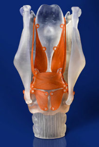 The structure of the laryngeal airway and arrangement and action of the vocal cords are often confusing to beginning anatomy students, especially using only two dimensional images. Significant faculty time and use of expensive anatomical models are needed to clarify these concepts. We are designing a simple, inexpensive, functional model of the larynx to be constructed through 3D printing as an interactive teaching and learning tool for health professions students at many levels and in numerous settings, e.g., self-study, flipped classroom, lab, and pre-exam review.
Assembling this simple model will combine cognitive and psychomotor skills and encourage students to transform kinesthetic experience into useful learning. We are also developing an educational SoftChalk package on the larynx to be used in conjunction with the 3D model for teaching beginning students in medicine, dentistry, and the health professions, as well as providing a review for advanced students, including residents and post-doctoral fellows. The package will include html5 and SCORM compatible content and formative assessment activities for self-study and in-class use via mobile devices. The model and educational package will be disseminated through educational sites such as MedEdPORTAL and the NIH 3D Print Exchange.
Supported by a grant from the University of Texas Kenneth I. Shine Academy of Health Science Education Small Grants Program.
The Anterior Abdominal Wall: A Self-Study Paper Model:
(Dr. Alan Sakaguchi, Dr. Maria Bartanuszova, Dr. Earlanda Williams, Dr. Charleen Moore)
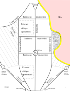 The anterior abdominal wall has numerous layers that are often confusing to beginning anatomy students. We have developed a simple, paper model of the anterior abdominal wall that is easily printed and constructed. Assembling this simple model of these complex anatomical structures is a fun and interactive way that encourages students to transform kinesthetic experience into useful learning. The model (of a male) uses colored paper or is printed in color to emphasize the multilayered structure of the abdominal wall. Each layer is labelled with some of the associated anatomical features that can be taught with the model.
Small, moveable cutout window flaps in the layers correctly show the aponeurotic composition of the laminae of the rectus sheath both anterior and posterior to the rectus abdominis muscle and above and below the arcuate line. The model incorporates superficial and deep inguinal rings, inferior epigastric vessels and the spermatic cord. As a result, the model can be used to teach the general structure of the inguinal canal, the fascial layers of the spermatic cord and where direct and indirect inguinal hernias would pass through the abdominal wall. An Instructor’s Guide and Assessment Questions have been developed to provide suggestions for using the model in lecture and laboratory settings as well as during peer-to-peer teaching, team-based learning and self-study.
The model has been published in MedEdPORTAL: Sakaguchi A, Bartanuszova M, Williams E, Moore C.
The Anterior Abdominal Wall: A Self-Study Paper Model. MedEdPORTAL; 2014.
Virtual Histology:
(Frank Weaker and Tom King)
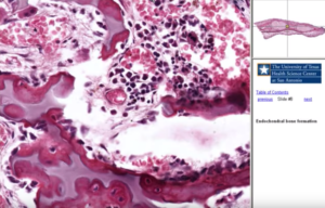 With the support of the SBC/ AT&T, Virtual Microscopy for the Health Profession (VMHP) was introduced to both Medical and Dental microscopic anatomy courses. (VMHP) is a comprehensive computer-based set of digitized histology and pathology color scanned images of actual human tissue specimens. Students can interact with and explore the specimen images in exactly the same fashion as on an actual specimen slide and light microscope.
Because these images are viewed using the student’s computer, the program is highly portable, user-friendly and convenient, i.e., it can be used whenever and wherever desired. The user can also “capture screen” as a tif- or jpeg-extension files. These image files can be sent electronically (e.g., e-mail attachments) from student to instructor for verification of cell or tissue identifications. These collected images could also be assembled into student atlases or practice practical examinations. The light microscope and glass slides are now a supplement to the VMHP.
GALEN III:
(Linda Johnson, Larry Roberman)
 The original software for the Galen program was written by Dr. Larry Roberman, one of our past medical school graduates. Current faculties have worked closely with Dr. Roberman to produce a question bank of over 2500 questions which have appeared on past examinations in Gross Anatomy and Embryology. Many of these questions are in clinical vignette format and duplicate the style of questions that appear on the USMLE Step 1 Exam. These questions can be called up for individual lectures, course modules, or on the basis of key words and can be used to self-test for both study and exam preparation. Students can mark difficult questions and return to them for future study. Students are given weekly Galen tests to encourage them to keep up with course material and identify weak areas in time for them to be remedied.
UT Health San Antonio Virtual for Medical Embryology Teaching:
(Kristine Vogel)
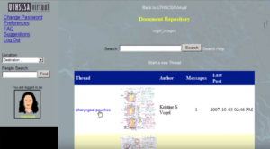 Embryology is intensely visual and involves dynamic terminology, which makes it a difficult topic for many medical students. The Medical Embryology Teaching tutorial was developed especially for those students who did not have a traditional undergraduate developmental biology course. UT Health SA Virtual is an online environment designed to facilitate academic collaboration, is accessible from any computer in the world with a current Internet browser, and is available to all UT Health SA faculty, staff, and students. Some of the options include virtual meetings with transcripts, a virtual reference librarian, an interactive shared desktop, secured document repository, and a personal office with image repository.
GATEways:
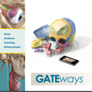 GATEways (Gross Anatomy Teaching Enhancement) project is a collaboration among faculty and staff of all six schools of UT Health SA to design, develop, and implement four technological initiatives to be used in teaching gross anatomy throughout the UT Health SA. Patricia Brewer (School of Allied Health and cross-appointed in CSBL) is the project leader, while other CSBL faculty serve as initiative leaders. GATEways has been supported by grants from the SBC/AT&T Foundation and local grants from the UT Health SA.
Dissection Video:
(Vick Williams)
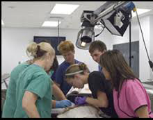
 To assist in the development of skill in manipulation of instruments and in following instructions for dissecting a cadaver, video demonstrations have been developed for instructional use and for reference by students. In addition to showing the anatomy of regions under study, these demonstrations are designed to emphasize technique. The videos are recorded in digital format that can simulate “high definition” and is available as whole laboratory units on DVD (for maximum resolution) video compact disk, from which individual segments of a dissection can be viewed in PowerPoint, or podcast.
Cranial Nerve Study Guide:
(Frank Weaker)
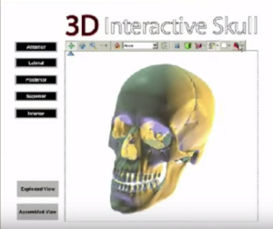 The study guide is a unique view of the twelve cranial nerves drawn into a three-dimensional skull. The Cranial Nerve Selector function allows the user to choose the individual nerve to study. Each cranial nerve module is divided into four sections. The Introduction is a narration of the fiber content, general function and branches of each nerve. The Animation is a narrative description and visualization of the pathway of the cranial nerve and its principal branches through a three-dimensional skull.
The Self-Directed section allows the student interactive control over a movie that traces the nerve. At various points, the student will be prompted to identify nerve branches and related structures. The Quiz section allows the students to answer questions about each module and to help measure their degree of understanding. The Advanced section is for the student seeking additional information of the fiber types that are found in each cranial. A tour of the tutorial can be viewed by the link provided below.
Anatomical Question Bank:
(Omid Rahimi and Linda Johnson)
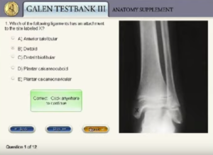 The Galen Test Bank II is being adapted into a new test bank that includes over 300 digital radiographic images and still photographs from dissection films and animations. The test bank will be available to faculty and students on the university server, on CD/DVD, incorporated into courses using Blackboard®, and in the classroom using the Turning Point Audience Response System®. A question from Galen Test Bank III is illustrated on the link provided below.
SkyEye® Camera Prosection Videos:
(Omid Rahimi)
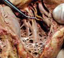
 The Sky-Eye® is a specially designed camera on a boon arm that allows a unique view of dissections. Dissections can be video captured and be broadcasted to classrooms and distant-learning sites.
Atlas of Human Coronal Brain Sections Stained by LeMasurier Method Labeled:
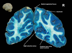 Atlas of Human Coronal Brain Sections Stained by LeMasurier Method Labeled
PowerPoint SlideShow – Created by: William W. Morgan, Ph.D.
Laboratory Software Development Projects:
Student familiarity with the usage of personal computers has mushroomed and this has been reflected in the greater use of computers at undergraduate universities around the nation. Several of the faculty in our department surveyed the software packages available for professional students at medical and dental schools and found it woefully lacking. A commitment was made by several of our course directors to produce our own educational software specific to the needs of our courses and students. The authors for these programs are shown along with the software titles, years in service, and link to a short virtual tour of the content. All programs are annually refined and updated and continue to be a success with our students.
Authors |
Title of CD-ROM |
Years in
|
|---|---|---|
|
Vaughan, M.K.
|
1997-2008
|
|
|
Weaker, F.J.,
Vaughan, M.K. |
1997-2008
|
|
|
Vaughan, M.K.
|
1998-2008
|
|
|
King, T.S.
Vaughan, M.K. & Weaker, F.J. |
1998-2008
|
|
|
Weaker, F.J. &
Vaughan, M.K. |
2000-2008
|
|
|
Vaughan, M.K. &
Johnson, L.Y. |
Medical Gross Anatomy X-Rays
|
2001-2004
|
|
Vaughan, M.K.
Johnson, L.Y. & Rahimi, O.B. |
2005-2008
|
|
|
Vaughan, M.K.
|
2001-2008
|
|
|
Vaughan, M.K.
|
2002, 2005
|